last authored: Aug 2016, Matthew M, Shivani Patel
Introduction
The heart is responsible for pumping oxygenated blood through the systemic circulatory system and deoxygenated blood to the lungs through the pulmonary circulatory system.

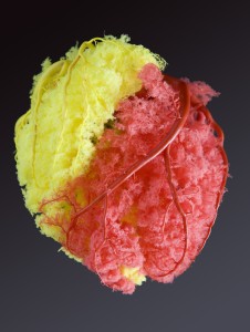
Coronary arteries; red = left; yellow = right; courtesy of Schandra
Due to the tremendous and sustained force the heart must exert during each beat, the heart consumes a large proportion of circulating oxygen. Although it counts for only 1/200 of the body’s weight, it requires almost 1/20 of total blood supply at rest.
The demand of supplying the heart with oxygen is met by a unique set of vessels known as the coronary arteries. These arteries are 5-10 cm long and 2-4 mm in diameter, surround the heart, and are filled with blood during the relaxation portion of the cardiac cycle.
The coronary veins and the coronary sinus into which they drain are then responsible for collecting deoxygenated blood and returning it back into circulation.
Major Arteries
The left and right coronary arteries leave the ascending aorta just after it emerges from the aortic valve. A posterior coronary artery may be present in some individuals, but not all.
The right coronary artery does not have major branches.
The left coronary artery, or left main artery, continues to divide into the left anterior descending (LAD) (also referred to as the anterior interventricular) and left circumflex (LCX).
Thus, the three major coronary arteries are the LAD, LCX, and right coronary artery (RCA).
Location
 The LAD is located in the anterior interventricular sulcus – a groove that separates the ventricles of the heart. It ends at the apex. One or more diagonal branches (D1, D2) leave the LAD.
Territories
The LAD supplies oxygenated blood to the majority of the left ventricular walls. Diagonal branches reach the anterior left ventricle, while septal branches reach the anterior septum. The LAD also supplies the AV node.
Location
 The LCX can be found in the coronary sulcus – a groove on the outer surface of the heart that marks the division between the atria and the ventricles.
Marginal branches (M1, M2, etc) leave the LCX. In most people the LCX ends in one of these marginal branches, but in 10% of people, it also supplies the posterior descending artery.
Territories
The LCX supplies the left atrial and ventricular walls of the heart with oxygenated blood.
The RCA continues in the AV groove posteriorly and gives off a branch to the AV node.
In 65% of cases the posterior descending artery (PDA) is a branch of the RCA (right dominant circulation).
The PDA supplies the inferior wall of the left ventricle and inferior part of the septum.
Location
 The RCA travels inferior to the right auricle, where it divides into two branches: the posterior interventricular and marginal branches.
The right coronary artery travels between the right artium and right ventricle and ends at the inferior part of the septum. The first conus branch reaches the R ventricular outflow tract. A large acute marginal branch runs along the margin of the R ventricle.
In 70% of patients, the Posterior Descending Artery (PDA) arises from the RCA. In 10% the PDA arises from the LCX, while in 20% it is supplied by both.
The RCA distributes oxygenated blood to the:
- right ventricle
- right atrium, through atrial branches
- SA node (in 60% of people)
- AV node (in 90% of people)
The PDA supplies the posterior third of the septum.
Smaller Arteries and Capillaries
Major vessels divide into epicardial arteries, while intramusclular arteries deviate to penetrate the myocardium perpendicularly to form subendocardial plexuses (Ramanathan &Skinner, 2005).
The heart also contains numerous, naturally formed anastomosis – sites where two or more arteries meet to supply oxygenated blood. These anastomoses allow for collateral circuits, in which blood can flow through more than one route to reach an area of tissue. Due to the existence of these anastomoses, the heart muscle can continue to be perfused even if one of the coronary arteries is blocked. (See Figure 4).
Coronary Veins

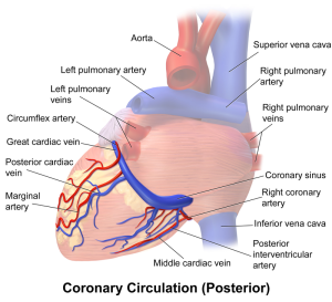
Coronary sinus, courtesy of Bruce Blaus
Most coronary veins return deoxygenated blood from the myocardium to the coronary sinus, located alongside the right atrium. From here, blood then returns to the general circulation.
The great cardiac vein is located anteriorly. It begins at the apex of the heart and ascends along with the LAD, then runs between the left atrium and ventricle before continuing as the coronary sinus. The posterior cardiac vein runs along the left ventricle, and drains into either the great cardiac vein or the coronary sinus. The middle cardiac vein is located posteriorly. It begins at the apex of the heart and ascends between the ventricles before draining into the coronary sinus. The small cardiac vein runs along the margin of the heart, then posteriorly in the coronary sulcus, draining into the coronary sinus. The anterior cardiac vein drains the anterior right ventricle, bypassing the coronary sinus and delivers blood directly into the right atrium.
Resources and References
Images provided by Robin Smithuis and Tineke Willems, Radiology Assistant


